Home/
Unlabelled
/Right Shoulder Anatomy Diagram - Muscles Of The Pectoral Girdle And Upper Limbs Anatomy And Physiology - This tool is at the same time useful for the training and teaching of the.
Right Shoulder Anatomy Diagram - Muscles Of The Pectoral Girdle And Upper Limbs Anatomy And Physiology - This tool is at the same time useful for the training and teaching of the.
Right Shoulder Anatomy Diagram - Muscles Of The Pectoral Girdle And Upper Limbs Anatomy And Physiology - This tool is at the same time useful for the training and teaching of the.. The muscles in the shoulder aid in a wide. We think this is the most useful anatomy picture that you need. This tool is at the same time useful for the training and teaching of the. On the anterior side of the shoulder, the coracobrachialis, serratus anterior, pectoralis major, and pectoralis minor muscles work as a group to flex and adduct the scapula and humerus anteriorly toward the sternum. Related posts of diagram of shoulder muscles and tendons muscle anatomy of the human body.
The muscles in the shoulder aid in a wide. Most people with rotator cuff injuries can recover with rest and physical therapy. These muscles form the outer shape of the shoulder and underarm. On the left is a standard (anatomic) shoulder arthroplasty. Deltoids anatomy when most people think of the
Human Anatomy Diagram Human Anatomy For Muscle Reproductive Skeletal Nerve Arm Muscle Anatomy Leg Anatomy Shoulder Muscle Anatomy from i.pinimg.com The most flexible joint in the entire human body, our shoulder joint is formed by the union of the humerus, the scapula (or shoulder blade), and the clavicle (or collarbone). Each anatomical structure was interactively labeled. This is the main muscle that lets you rotate and extend your shoulder. We'll go over the bones, joints, muscles, nerves, and blood vessels that make up the human arm. The shoulder is a complex combination of bones and joints where many muscles act to provide the widest range of motion of any part of the body. This tool is at the same time useful for the training and teaching of the. Contents hide 1 anatomical terms. The muscles in the shoulder aid in a wide.
A second joint in the shoulder is the junction of the collar bone with the shoulder blade.
The shoulder is made up of two joints, the acromioclavicular joint and the glenohumeral joint. On the anterior side of the shoulder, the coracobrachialis, serratus anterior, pectoralis major, and pectoralis minor muscles work as a group to flex and adduct the scapula and humerus anteriorly toward the sternum. Plus, exercises for training them. We'll go over the bones, joints, muscles, nerves, and blood vessels that make up the human arm. A person bends the elbow 90 degrees (at a right angle) while gripping hands with the health care professional, who applies pressure to the arm. Each anatomical structure was interactively labeled. Many in the neck help to stabilize or move the head. What does a torn shoulder labrum feel like? The glenohumeral joint is where the ball (humeral head) and the socket (the glenoid) meet. However, more serious injuries, such as complete rotator cuff tears, may require surgical repair. These muscles form the outer shape of the shoulder and underarm. This is the main muscle that lets you rotate and extend your shoulder. A diagram of an anatomic shoulder replacement—the plastic socket replaces the cup of the scapula (shoulder blade).
The acromioclavicular joint is where the acromion, part of the shoulder blade (scapula) and the collar bone (clavicle) meet. Numerous muscles help stabilize the three joints of. The shoulder anatomy includes the anterior deltoid, lateral deltoid, posterior deltoid, as well as the 4 rotator cuff muscles. The arm is one of the body's most complex and frequently used structures. On the anterior side of the shoulder, the coracobrachialis, serratus anterior, pectoralis major, and pectoralis minor muscles work as a group to flex and adduct the scapula and humerus anteriorly toward the sternum.
E153 Shoulder Anatomy Shoulder Blade Muscles Youtube from i.ytimg.com On the left is a standard (anatomic) shoulder arthroplasty. The acromioclavicular joint is where the acromion, part of the shoulder blade (scapula) and the collar bone (clavicle) meet. Anatomynote.com found right arm muscle and tendon anatomy from plenty of anatomical pictures on the internet. The components of the ball and cup are reversed on the right—a reverse shoulder replacement. Elbow fractures icons orthopedic impingement body yoga anatomy back shoulder elbow fracture glenoid icons pain shoulder and elbow pain shoulder joint. Pain in a specific shoulder area. Looking for quizzes, videos, articles and an atl. It helps you make all the motions of your arm and shoulder.
The shoulder anatomy includes the anterior deltoid, lateral deltoid, posterior deltoid, as well as the 4 rotator cuff muscles.
This tool is at the same time useful for the training and teaching of the. 2.2 shoulder muscles and shoulder tendons. On the left is a standard (anatomic) shoulder arthroplasty. New york injury cases blog these pictures of this page are about:right shoulder anatomy. The shoulder is a complex combination of bones and joints where many muscles act to provide the widest range of motion of any part of the body. Contents hide 1 anatomical terms. Anatomynote.com found right arm muscle and tendon anatomy from plenty of anatomical pictures on the internet. This is the main muscle that lets you rotate and extend your shoulder. Superficial muscles are the muscles closest to the skin surface and can usually be seen while a body is performing actions. The acromioclavicular joint is where the acromion, part of the shoulder blade (scapula) and the collar bone (clavicle) meet. This is the smallest rotator cuff muscle. The shoulder is made up of two joints, the acromioclavicular joint and the glenohumeral joint. Plus, exercises for training them.
We'll go over the bones, joints, muscles, nerves, and blood vessels that make up the human arm. Numerous muscles help stabilize the three joints of. However, more serious injuries, such as complete rotator cuff tears, may require surgical repair. Each anatomical structure was interactively labeled. The shoulder joint is the junction between the chest and the upper extremity.
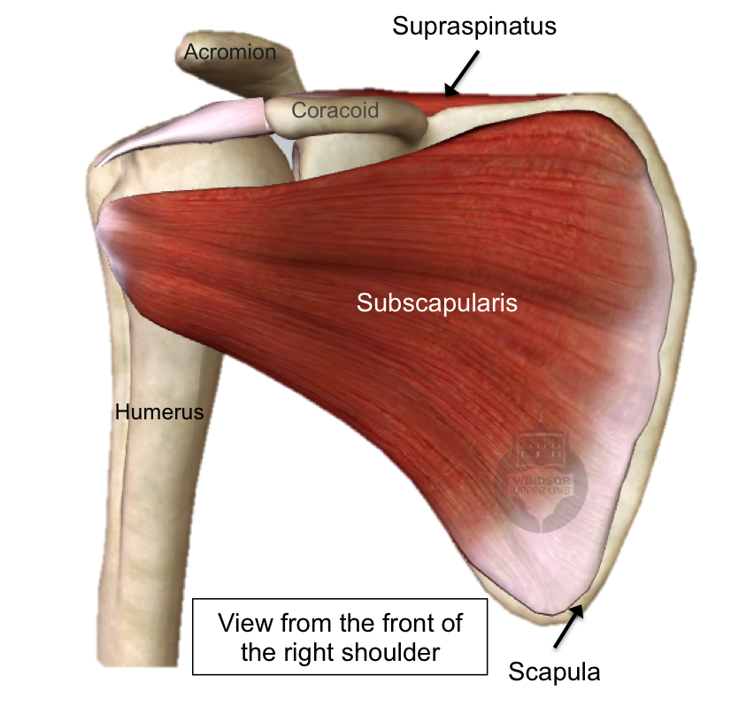
Rotator Cuff Tear from www.windsorupperlimb.com The anatomy of the shoulder. It helps you make all the motions of your arm and shoulder. Pain in a specific shoulder area. The components of the ball and cup are reversed on the right—a reverse shoulder replacement. Located superior to the shoulder joint, the deltoid muscle works with the supraspinatus to abduct the arm at the shoulder. The primary function of the shoulder girdle is to give strength and range of motion to the arm. Elbow fractures icons orthopedic impingement body yoga anatomy back shoulder elbow fracture glenoid icons pain shoulder and elbow pain shoulder joint. Sechrest, md narrates an animated tutorial on the basic anatomy of the shoulder.
Anatomy of the shoulder interesting facts about shoulder anatomy.
Numerous muscles help stabilize the three joints of. Ebraheim's educational animated video describes muscle anatomy of the shoulder girdle and anatomy of the shoulder joint.anatomy of the shoulder muscles a. In this episode of eorthopodtv, orthopaedic surgeon randale c. Updated on may 26, 2020. These symptoms may vary depending on the type of labral tear a person has. Most people with rotator cuff injuries can recover with rest and physical therapy. See shoulder anatomy stock video clips. Looking for quizzes, videos, articles and an atl. Many in the neck help to stabilize or move the head. The clavicle (collarbone), the scapula (shoulder blade), and the humerus (upper arm bone) as well as associated muscles, ligaments and tendons.all together it is made up of four joints as well as the muscles that are responsible for movement in the shoulder attached to the scapula. The muscles in the shoulder aid in a wide. A diagram of an anatomic shoulder replacement—the plastic socket replaces the cup of the scapula (shoulder blade). We'll go over the bones, joints, muscles, nerves, and blood vessels that make up the human arm.
However, more serious injuries, such as complete rotator cuff tears, may require surgical repair shoulder anatomy diagram. The most flexible joint in the entire human body, our shoulder joint is formed by the union of the humerus, the scapula (or shoulder blade), and the clavicle (or collarbone).
Right Shoulder Anatomy Diagram - Muscles Of The Pectoral Girdle And Upper Limbs Anatomy And Physiology - This tool is at the same time useful for the training and teaching of the.
Reviewed by WAN waN
on
April 26, 2021
Rating: 5


 Reviewed by WAN waN
on
April 26, 2021
Rating:
Reviewed by WAN waN
on
April 26, 2021
Rating:

Post a Comment